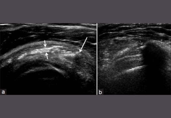
Calcific bursitis of the shoulder
Inflammation of the bursa can lead to calcium deposition most commonly seen within the sub-acromial bursa of the shoulder.

Subjective History
Often pain felt at the acromion
Pain may radiate to upper arm
Pain may be felt at rest and often worse at night
Patient may report a history of, gradual onset, repetitive upper limb activity; history of rotator cuff trauma
Often aggravated by shoulder abduction
Objective Examination
Positive painful arc – pain between 60 and 120 degrees
It is important to note that calcific bursitis and a rotator cuff pathology can co-exist
Limited active range secondary to pain
Full passive range of motion
Ultrasound/ X-ray or MRI can be used to confirm diagnosis
References
Image from OpenI – Licensed by CC
Treatment
0-48 hours NSAID’s, ice , rest
Physiotherapists may apply tape in the acute stage to help with a patients perception of pain and provide symptom relief
After 48 hours encourage active or active assisted shoulder range of movement within pain-free range
Once pain has subsided a rotator cuff strengthening programme can be implemented
For chronic persistent symptoms a corticosteroid injection under ultrasound guidance may be considered
References
Image from OpenI – Licensed by CC


